- Lecture
On October 10, 2026, at 3:00 PM
(L-18) Development of an AI-based 3D scanning method for generating a 3D-printed, silicone-based epithesis in oral and maxillofacial surgery
Speaker: Univ.-Prof. Dr. Dr. Ralf Smeets I Hamburg, DE
Summary:
Objective:
The treatment of patients with facial defects, such as those caused by congenital malformations, presents a particular challenge not only for an interdisciplinary treatment team—comprising oral and maxillofacial surgeons and prosthodontists—but also for the patients themselves. When congenital or acquired defects in the head and neck region cannot be treated through surgical intervention—particularly following cancer or trauma—the existing defect must be reconstructed using prostheses. These prostheses not only conceal the defect but are also intended to be aesthetically sophisticated, thereby facilitating the patient’s psychosocial reintegration. Such an epithesis often represents the last chance for patients, for example, following chemotherapy. The working group’s goal was to capture and convert suitable data using AI to generate a detailed, lifelike 3D image for epithesis modeling.
Materials/Methods:
Various 3D scanning technologies were investigated to identify the most suitable method for rapid data acquisition without ionizing radiation. The target scan time was 30–60 seconds with a maximum radiation exposure of 200 μSv. A protocol to support data acquisition and ensure sufficient image quality was developed in collaboration with the project partners. This includes performing scans in a dedicated room with optimal lighting conditions, maintaining sufficient distance from the patient during the scanning process, and minimizing patient movement. Additionally, a method was developed to generate a 3D depth map of the scanned area, which enabled subsequent modeling.
Results:
Initial tests with selected technologies have shown that the target data acquisition time is achievable. Radiation exposure remains well below the limit. Point accuracy: The required point accuracy of at least 100 μm for capturing fine textural details, such as pores and/or wrinkles, has been successfully validated. Methods are currently being developed to accurately capture fine texture details on the surface of the face and neck, including the correct representation of cavities in the 3D model. The ideal peripheral lighting according to CIE Standard D65 was investigated. This revealed that under “standard clinical lighting” in the examination room, without special lighting measures, the defect scan can be performed with sufficient accuracy. A color depth of at least 24 bits per pixel was determined to be necessary to precisely map realistic skin discoloration. This parameter can be technically achieved within the framework of modern scanning methods. Using AI algorithms, we have developed and successfully implemented algorithms for noise filtering, gap filling, and normalization of the captured data. These steps are crucial for bringing the data into a uniform coordinate system and improving quality.
Conclusion:
This project focuses on automating the workflow for capturing and processing 3D data. So far, we have established a stable pipeline; however, there are still challenges in integrating user-friendly interfaces to make the system’s complexity more accessible to non-technical users. One of the biggest challenges lies in the quality of the captured data, which is often compromised by noise and artifacts. Additionally, processing large point clouds requires significant computational resources, which can lead to longer processing times. Developing a user-friendly interface that makes the complex functions accessible is also a challenge.
Conflict of Interest: We thank the Federal Ministry for Economic Affairs and Climate Action for its financial support (Grant No.: KK5180710IE3).
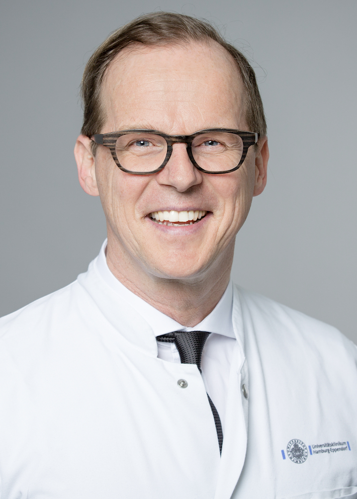
Speaker:
- Univ.-Prof. Dr. Dr. R. Smeets studied Chemistry from 1990 to 1995, specializing in Macromolecular Chemistry during his main course of study. He subsequently studied Human Medicine and Dentistry at RWTH Aachen University from 1995 to 2003. In 2004 and 2005, he received a research fellowship from RWTH Aachen University.
- In 2008, Prof. Smeets obtained his specialist qualification in Oral and Maxillofacial Surgery, followed by specialist certification in Oral Surgery in 2009. In recognition of his scientific and clinical achievements, he received the Hans von Seemen Prize from the German Society for Plastic and Reconstructive Surgery in 2010. In 2011, he was appointed Managing Senior Physician and Head of Research at the Department of Oral and Maxillofacial Surgery at the University Medical Center Hamburg-Eppendorf (UKE). In the same year, he was appointed W2 Professor of Oral and Maxillofacial Surgery and Oral Surgery at the Faculty of Medicine of the University of Hamburg.
- From 2012 to 2015, Prof. Smeets served as a visiting lecturer at Bremerhaven University. Within the German Society of Dentistry and Oral and Maxillofacial Medicine (DGZMK), he held various positions in scientific working groups. From 2012 to 2014, he served as Secretary of the Working Group for Basic Research (AfG), and from 2013 to 2015 as Secretary of the Working Group for Oral Pathology (AKOPOM). From 2014 to 2016, he was President of the AfG. Since 2015, he has also served as Vice Chair of AKOPOM.
- Since 2017, Prof. Smeets has been a member of the Board and Head of Continuing Education of the German Association of Oral Implantology (DGOI). From 2018 to 2023, he held a Heisenberg Professorship funded by the German Research Foundation (DFG) in the field of “Regenerative Orofacial Medicine.” Since 2018, he has also held a W3 Professorship in Oral and Maxillofacial Surgery and “Regenerative Orofacial Medicine” at the Department of Oral and Maxillofacial Surgery at the University Medical Center Hamburg-Eppendorf. In the same year, he became Head of the independent Section of “Regenerative Orofacial Medicine.” He was also elected a Fellow of the International Team for Implantology (ITI Fellow) in 2018.
- From 2019 to 2024, Prof. Smeets served as Deputy Director of the Department of Oral and Maxillofacial Surgery at the University Medical Center Hamburg-Eppendorf. Since 2020, he has been a Visiting Professor at the Department of Oral and Maxillofacial Surgery at Hebei Medical University in Shijiazhuang, People’s Republic of China. Since 2021, he has served as an advisor to the Fraunhofer IAPT in the field of medical applications for the head and neck region, with a particular focus on medical technology. In the same year, he became a member of the Board of the DFG Research Group 5250.
- Since 2022, Prof. Smeets has also served as a visiting lecturer at TU Dortmund University and as a member of the Board of the University Center for 3D Printing in Medicine and Pharmacy (UC3D). Since 2022, he has additionally held the position of Vice President and Head of Continuing Education of the German Association of Oral Implantology (DGOI).
- Prof. Smeets has received numerous awards in recognition of his outstanding scientific, clinical, and educational achievements. These include the German Wound Award 2022, the German Hygiene Award 2022 presented by the Rudolf Schülke Foundation, and the Hamburg Teaching Award 2022, which was presented in 2023.
- Prof. Smeets has extensive scientific expertise and a strong track record in national and international research, teaching, and academic lecturing. His scientific work comprises more than 285 publications, including more than 260 articles in peer-reviewed journals. He is also the holder of several patents and has received numerous additional awards. His academic profile is complemented by extensive national and international activities as a lecturer, invited speaker, and peer reviewer.
Facility / Institute / Clinic:
- Department of Oral and Maxillofacial Surgery
- Section for Regenerative Orofacial Medicine
- University Medical Center Hamburg-Eppendorf
- Martinistraße 52
- 20246 Hamburg, DE
Co-authors:
- Maison, W., Fuest, S., Gosau, M., Voos, A.
- Kraume, J., Weber, V., Wolf, N., Sauer, T.
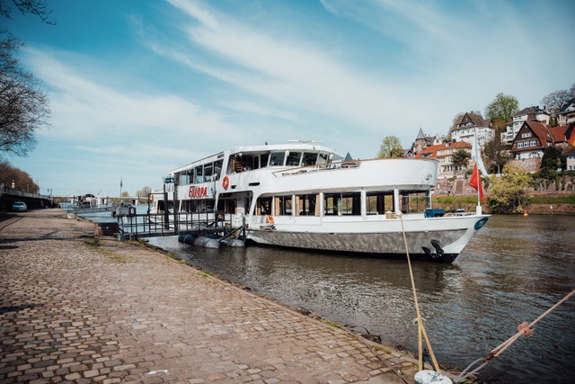
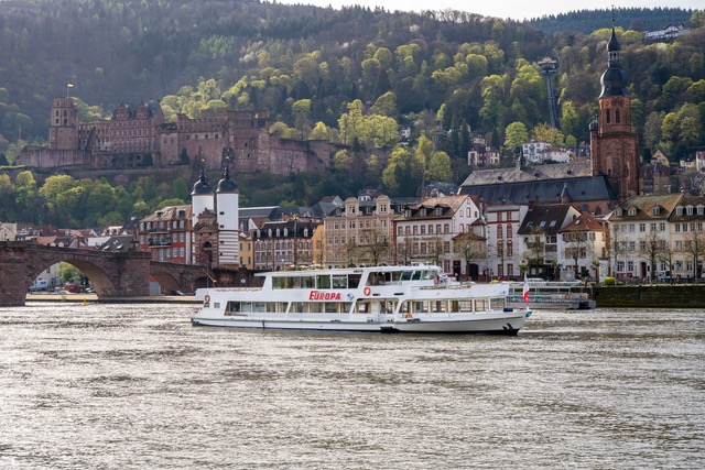
Shipping “Weisse Flotte”
At the end of the congress, we will enjoy an enjoyable evening together on board of the “Weisse Flotte”.
Only a few tickets left